

- 발행인: 박인양
- 편집인: 이경아
- 발행: 대한산부인과초음파학회
증례
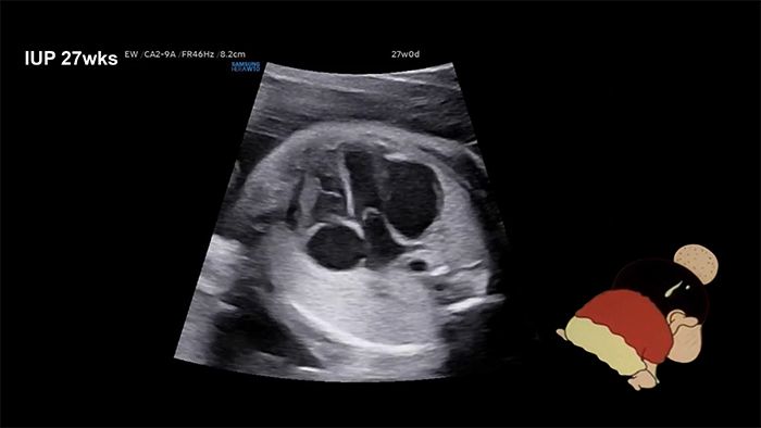
A 36-year-old woman was referred at 27 weeks of gestation for evaluation of a suspected dysplastic mitral valve (MV) with mitral regurgitation (MR). Ultrasonography revealed a 20 x 14 mm abnormally dilated, pouch-like lesion adjacent to the left ventricle (LV), with minimal blood flow into the pouch, and associated severe MR.
LV aneurysm or diverticulum was suspected. No signs of fetal hydrops
were observed. Despite the pouch gradually enlarging as gestation progressed, the fetus remained stable.
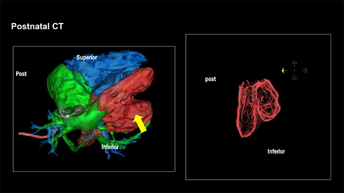
At 39.5 weeks of gestation, a cesarean section was performed, delivering a female infant weighing 3,750 g. Postnatal echocardiography and cardiac computed tomography confirmed a diagnosis of LV diverticulum with an MV anomaly.
The diverticulum extended from the base of the LV to approximately two-thirds of its length. The baby is stable and awaiting corrective surgery.
Keywords: LV diverticulum